




Class Roster








































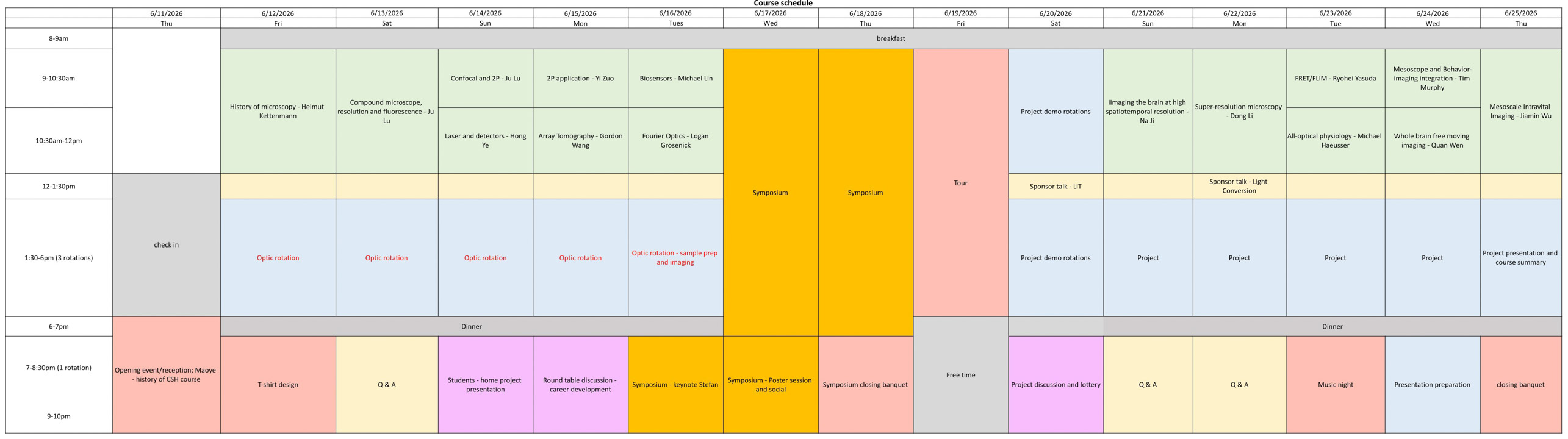
Schedule
Projects
During the second week of the course, students conduct hands-on research projects led by faculty members using state-of-the-art imaging systems.
Multi-region neuronal imaging in freely behaving mice with TINIscope
Students will learn to image neuronal activity from multiple brain regions of freely moving mice using 0.43g head-mounted TINIscopes. They will compare neuronal dynamics and interregional coordination across cortical and subcortical circuits during different behavioral tasks. The project covers multi-region data acquisition, behavioral tracking, and alignment of neural activity with behavior to explore large-scale circuit functions.
Integration of Miniature 2-photon Imaging with Behavioral Monitoring
This project introduces students to a complete experimental workflow linking neural activity in the medial prefrontal cortex (mPFC) to freely moving mouse behavior. Students will acquire high-quality 3D video recordings of mice during tasks, analyze their movement, and explore how to integrate behavioral and neural data.
Mesoscale Intravital Fluorescence Microscopy of Neural Activity
Students will use a mesoscale intravital fluorescence microscope to capture neural activity across broad brain regions in awake mice. The goal is to record population-level neuronal responses at single-cell resolution across extended cortical regions in response to diverse visual stimuli.
All-Optical Circuit Interrogation Using Targeted Photostimulation
Students will first image cortical neurons to identify those that respond to given sensory stimuli. Then, by targeted two-photon photostimulation of those identified neurons, they will attempt to activate defined ensembles. Simultaneously, calcium imaging will be used to monitor responses in downstream “follower” neurons, enabling investigation of circuit connectivity.
Fluorescence Lifetime Imaging Microscopy (FLIM) Photometry
In this project, students will learn how to perform in vivo FLIM-based fiber photometry to record neuronal activation or neuromodulatory signals using fluorescent biosensors in freely behaving animals. The project will provide hands-on experience with cutting-edge functional imaging approaches, including both data acquisition and FLIM data analysis. This training will help students develop practical skills in experimental design, optical recording, behavioral neuroscience, and quantitative analysis of functional imaging data.
Light Sheet Microscope for Large Scale In Vivo and In Vitro Imaging
Students in this project will get hands-on experience operating a light-sheet microscope to image large volumes — either whole cleared mouse brains or living zebrafish. With live imaging in fish, we will capture neuronal activity, immune cell migration, and other fluorescently labeled biological dynamics within intact organisms. Students will also learn data analysis workflows for large 3D datasets.
Chronic Monitoring of Single-Synapse Transmission During Associative Learning
Students will use a state-of-the-art genetically encoded glutamate sensor (iGluSnFR) together with an optimized two-photon microscope for longitudinal imaging. The goal is to directly visualize glutamate release events at individual synapses in awake, behaving animals. This project aims to build a complete “activity history” of individual synapses over the course of learning.
Correlative In Vivo Imaging with Terminal Analysis at Super-resolution (CIVITAS)
In this project, students will combine in vivo two-photon imaging with post-mortem expansion microscopy and super-resolution STED imaging to link structural, functional, and molecular information at single-synapse resolution. Specifically, cortical dendrites in live mouse cortex will first be imaged with the 2P microscope. After perfusion and fixation, the same dendrites will be re-identified in the fixed brain tissue.
Sponsors











Fun & Photo Gallery
Capture and share course moments live on our PhotoWall. Tap below to upload photos and view moments shared by course participants.